 Per Drs. Rod Jackson, Joanna Broad, Jennie Connor, and Susan Wells of the University of Auckland, “Do not assume there is a window in which the health benefits of alcohol are greater than the harms…” [1].
It is common to hear conventionally-trained cardiologists and other healthcare providers recommend a reasonable intake of wine or state that a periodic consumption of alcohol can actually help prevent against the onset of cardiovascular disease. Unfortunately, the notion of alcohol being beneficial for the heart is misguided and dangerous, and in this short article I’ll explain why. Firstly, the majority of evidence that seemingly supports a low level of alcohol being in the diet comes from epidemiological or observational studies, and as Jayasekara et al. have stated, “The quality of the epidemiologic evidence relating to a protective effect of low-dose alcohol consumption has been challenged” [2]. Much of the impact of alcohol drinking on mortality and the progression of disease states has been misinterpreted or viewed narrow-mindedly, as a large meta-analysis published in 2014 has explained: “These findings suggest that reductions of alcohol consumption, even for light to moderate drinkers, may be beneficial for cardiovascular health. Our results therefore challenge the concept of a cardioprotective effect associated with light to moderate alcohol consumption reported in observational studies and suggest that this effect may have been due to residual confounding or selection bias” [3]. Uncontrolled confounding can account for at least some, if not all, of the reported cardioprotective associations in non-randomized studies, as concluded in a paper by Naimi et al., “These findings suggest that some or all of the apparent protective effect of moderate alcohol consumption on CVD may be due to residual or unmeasured confounding” [4]. The results of observational studies can also be distorted by underlying poor health in nondrinkers and the existence of greater underlying health in light and moderate drinkers versus heavy drinkers. Further corroboration has been offered by Shaper, Wannamethee, and Walker: “The data suggest that the observed alcohol-mortality relationships are produced by pre-existing disease and by the movement of men with such disease into non-drinking or occasional-drinking categories. The concept of a “protective” effect of drinking on mortality, ignoring the dynamic relationship between ill-health and drinking behaviour, is likely to be ill founded” [5]. We must also refrain from looking at variables in isolation, for the effect of alcohol on HDL (high-density lipoprotein) cholesterol and platelet aggregation can only be considered positive if we ignore all of alcohol’s negative effects on the cardiovascular system and the body at large [6]. In that vein, let’s now examine the physiological consequences of tippling. Moderate alcohol intake can increase plasma triglyceride concentration which can heighten cardiovascular disease risk in men and women independent of HDL cholesterol changes [7] [8]. Normally, plasma triglycerides are hydrolyzed by the enzyme lipoprotein lipase in capillary endothelial cells, but alcohol can inhibit this enzyme and keep plasma triglycerides elevated by hindering the typical maturation of VLDLs (very-low-density lipoproteins) to IDLs (intermediate-density lipoproteins) and then to LDLs (low-density lipoproteins) as triglycerides are removed by lipoprotein lipase [9]. Thus, the consumption of alcohol can boost the plasma’s concentration of triglyceride-rich lipoproteins which can be atherogenic (TGRLs can worsen arterial wall permeability and cause vascular injury) [10] [11]. Alcohol can also inhibit the secretion of VLDLs from the liver (promoting the development of fatty liver) by impairing hepatocyte microtubule formation or obstructing the synthesis of phosphatidylcholine [12]. Ideally, the secretion of VLDLs by the liver is adequate to thwart fatty liver disease but insufficient to cause an abnormal accumulation of triglycerides in the blood by way of overwhelming the circulatory system’s metabolizing of these lipid carriers. As a far better alternative to liquor, daily supplementation with niacin (vitamin B3) can greatly and safely drop plasma triglycerides and LDLs as well as raise HDLs [13]. In fact, niacin, as expressed by Carlson, “lowers the levels of all atherogenic lipoproteins – VLDL and LDL with subclasses as well as Lp(a) – and in addition it raises more than any other drug the levels of the protective HDL lipoproteins” [14]. Overarchingly, alcohol interferes with the normal metabolism of lipids in the body, and through multiple pathways this can encourage the development of cardiovascular disease [15]. Moving on to a comparison between grapes and wine, whole juice from purple grapes has been shown to decrease platelet aggregation, boost platelet-derived nitric oxide release, and lower superoxide (a strong free radical) production in vivo [16]. And red grape juice has been found to more effectively curb atherosclerosis and improve lipid and antioxidant parameters versus red wine or dealcoholized red wine [17]. Accordingly, we can attribute the genuine benefit to heart health from wine to the polyphenols found in grapes, and not to ethanol (ethanol can be directly toxic to the cardiovascular system and can raise blood pressure) [18]. Furthermore, resveratrol consumption through grapes but not through wine has been associated with a reduced breast cancer risk, while a heightened breast cancer risk has been associated with consuming red wine [19] [20]. Even a light intake of alcohol (less than or equal to one drink per day) can notably escalate oral cancer risk [21]. Furthermore, persistent wine intake can lead to dental erosion, and there is concern over the amount of toxic metals (like lead, cadmium, and arsenic) that can be found in various wines [22] [23]. Now there is no doubt that resveratrol itself can be greatly cardioprotective and beneficial to the body. Though it’s important to remember that resveratrol certainly is not the only acting polyphenol in wine and grapes [24]. Resveratrol can excellently exert anti-inflammatory, antioxidant, and vasodilation effects, in addition to being able to suppress tumor growth and protect neurons in the brain against damage [25] [26] [27]. More specifically, resveratrol helps to regulate redox homeostasis in humans and can block the oxidation of LDLs (drinking wine or other alcoholic beverages can up LDL oxidation), as well as induce apoptosis in cancer cells and better both insulin sensitivity and blood pressure [28] [29] [30] [31] [32]. After the ingestion of an alcoholic beverage, most of the ethanol will be oxidized in the liver by the enzyme alcohol dehydrogenase, yielding the highly toxic metabolite acetaldehyde [33]. Ideally, yielded acetaldehyde will be quickly converted into acetate by another liver enzyme, acetaldehyde dehydrogenase, with the help of glutathione. Acetate should then be converted into acetyl coenzyme A and ultimately into water and carbon dioxide. With a deficient supply of glutathione however, acetaldehyde can accumulate in the body and wreak havoc as it is a major generator of free radicals and a known carcinogen [34]. Also note that acetaldehyde occurs naturally in alcoholic beverages in addition to arising from ethanol metabolism [35]. We know that acetaldehyde directly impairs the contractile function of the heart, disrupts cardiac excitation-contraction coupling, and fosters lipid peroxidation and oxidative damage in the heart [36]. And ethanol metabolites termed ‘fatty acid ethyl esters’ can encourage injury to the liver, heart, and pancreas [37]. Acetaldehyde can also induce mitochondrial dysfunction and stress endoplasmic reticula, as well as negatively modify and form adducts with various proteins that stimulates the crafting of antibodies against acetaldehyde epitopes (which can lead to autoimmune responses) [38] [39] [40]. Acetaldehyde may contribute to organ damage (including the brain) by way of inflammatory cytokines and the same forming of protein adducts just mentioned too [41]. As a side note, acetaldehyde-induced dysfunction of the heart can be somewhat ameliorated by thiamine (vitamin B1) and folate (vitamin B9) [42]. Wrapping up my argument with a few more consequences of tippling, alcoholic beverages can suppress impeding wave motility in the small intestine (cutting the amount of time ingested food has to be properly digested), injure the gut mucosa and intestinal microbiota (potentially causing small intestinal bacterial overgrowth), magnify the translocation of bacterial toxins (like lipopolysaccharide) from the gut to the blood (which can contribute to liver and heart disease progression), and increase the permeability of the intestinal wall (creating a leaky gut) [43] [44] [45] [46] [47]. Importantly, ethanol (a carcinogen itself) can also facilitate the activation of procarcinogens (turning them into carcinogens) and the conversion of xenobiotics to toxic metabolites, enhancing the harmfulness of dietary and environmental toxins [48] [49]. Lastly, ethanol exposure can lead to atrophy of all skeletal muscle fiber types, lowered muscle capillarity, and altered metabolism in muscle tissue too [50]. In conclusion, I am not stating that alcohol, especially red wine, is the worst thing we could possibly put into our bodies. If you’d like to indulge in a drink here and there, by all means do your thing. Just please don’t assume that by knocking a few back you’re doing something to improve the health of your heart as some who should know better have suggested. Alternatively, simply eating some grapes and berries (plus some niacin-rich foods like chicken, turkey, sunflower seeds, green peas, and avocados) will beautifully supply your body with plenty to support your cardiovascular system and more without any of the ill effects of ethanol and its metabolites. Appropriately supplementing with isolated niacin and resveratrol would be another option for safely profiting the heart. Chalk another one up for food as medicine. References:
0 Comments
 For ages the salamanders have been considered a class of the elementals – vehicles of the transmuting fire element which they guard and embody. Dwelling in their subtle ether, these transubstantial nature spirits were often invoked and honored via the burning of incense. Later being associated with the element nitrogen as understood within modern chemistry, the salamanders are said to be operant in the emotions of man through the liver and bloodstream (the liver is believed to be the seat of the emotional principle and assists in processing emotions in addition to its biochemical transformation roles) [1]. Salamanders (as elementals) have been assigned the cardinal direction of south, and it is maintained that they are the most powerful of the nature spirits.
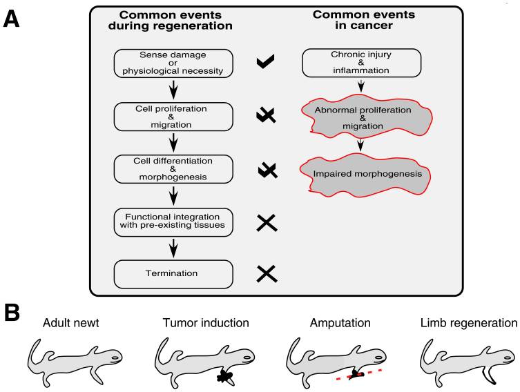
Salamanders are quite unique among vertebrates in their capacity to resist the development of cancer and regenerate limbs, and for many decades the link between regenerative ability and cancer protection has been discussed [2]. Normal tissue regeneration involves the directing of progenitor cells to an area of need, the careful proliferation and differentiation of those cells, and timely termination signals [3]. Aberrant or unregulated restoration efforts would obviously create unwanted consequences, but appropriate regeneration can actually halt and reverse the autonomous growth of malignant cells [4] [5] [6]. Because of the corollaries that exist between the events of tissue regeneration and the progression of cancerous growths, local tumors can be described as wounds that do not heal [7]. Chronic tissue stress or injury can drive ongoing inflammation which leads to an incomplete repair and restore operation that involves the misappropriating of mechanisms governing tissue mending and stem cell self-renewal – and this subversion fosters tumor maturation [8]. Because there are intracellular and extracellular factors that can regulate the self-renewal of stem cells, remedying the cancer pathology must include a reestablishment of normalcy and health to the microenvironment or terrain of the malignancy [9]. The remainder of this article will revolve around this concept as we look deeper into cancer’s idiosyncrasies. The tumor-suppressing protein p53 is frequently inactivated by mutations or viral oncogenes in human cancers, and Villiard et al. have shown that p53 signaling is necessary for limb regeneration in salamanders [10] [11]. And a related study provided evidence that more robust p53 proteins and a greater number of them confers heightened tumor resistance [12]. Ordinarily, p53 activation triggers DNA repair, apoptosis (mainly through the mitochondrial death pathway), and senescence signaling which helps to eliminate faulty cells and thwart cancerous growths [13]. Additionally, the p53 protein normally checks the self-renewal of adult stem cells, so when this protein is dysfunctional or inactive, stem cells will have less of a controlling leash, and therefore may abnormally proliferate more easily [14]. Again, the events of regeneration and tumorigenesis can be seen as two different but related paths. When the p53 protein is functioning as it should, it blocks the initial steps of regeneration from shifting toward tumorigenesis [15]. Thus, correct p53 activity helps prevent the transforming of healthy tissue restoration into unhealthy tissue malignancy. Moving forward, well-orchestrated sequences are necessary for the molding of replacement tissue in adult vertebrates to prohibit errant growth of mature cells that have dedifferentiated to fuel healing efforts. Salamanders skillfully employ dedifferentiation and transdifferentiation of mature cells to rebuild tissues that have been damaged or removed [16]. As Oviedo and Beane have explained, “in mammals chronic epithelial injury often precedes malignant transformation, while in urodeles (salamanders) and planarians (flatworms) persistent damage generally ends merely with functional repair. Injury response always entails attempts to repair the damage, but the fundamental differences probably lay in the coordination of such responses rather than in structural or species-specific capabilities” [17]. And evidence suggests that it is “individuation” or regenerative fields that direct and coordinate epimorphic regeneration in animals – a notion put forth by Conrad Hal Waddington in 1935 [18]. In 1938, Burr, Smith, and Strong found that bioelectric differences exist between cancer-susceptible and cancer-resistant mice, as well as that the onset of cancer is associated with a global deviation in an organism’s electrodynamic field [19]. Accordingly, we can state that cancer’s pathology is not merely the result of a random somatic mutation, and that it indeed consists of informational as well as molecular elements. Continuing, remodeling of the extracellular matrix is a major piece of limb regeneration and as a side note this process can be augmented with sufficient vitamin A [20]. Fibroblasts are prototypical cells that construct extracellular matrices and play central roles in both wound healing and tumor formation [21]. In salamanders, the dedifferentiation of fibroblasts (as well as some other cell types) after injury gives rise to what is known as a regeneration blastema (a mass of undifferentiated cells primed to repair lost tissue) that supplies the injured area with restorative cells [22]. In humans, the wound healing process can be divided into four phases: hemostasis, inflammation, proliferation, and tissue remodeling [23]. As the proliferation phase concludes in humans, certain fibroblasts (myofibroblasts) undergo apoptosis and are then cleared by macrophages [24]. In cancer, instead of being cleared away by macrophages, myofibroblasts remain in the diseased or injured region and become cancer-associated fibroblasts that are persistently activated by and promote the expansion of the developing tumor [25]. Now, earlier I mentioned the importance of reestablishing normalcy and health to the microenvironment of a tumor in the reversing of cancer because communication between neoplastic cells and their microenvironment drives tumor progression [26]. For corroboration, Bissell and Hines have explained that “the microenvironment can provide crucial signaling to maintain tissue architecture, inhibit cell growth and suppress or revert the malignant phenotype…incorrect signals from the microenvironment should lead to destabilization of tissue homeostasis and initiation and promotion of normal cells to malignancy” [27]. Salamanders masterfully create embryonic-like microenvironments at the site of regeneration after injury, and this not only ensures the correct reconstructing of harmed or missing tissue, but can also reprogram cancer cells (via signaling pathways) so that they revert to a normal phenotype [28]. Such reprogramming can also take place in the human body, and the homeobox genes (which code for proteins that help direct morphogenesis) that salamanders utilize in regeneration are also present in humans [29] [30]. Appropriate activation of beta-catenin and wnt signaling could trigger expression of the above genes that leads to commands and instructions for regeneration being given in Homo sapiens [31]. Alternatively, virus-mediated transduction could be used to extract functional homeobox genes from salamanders and insert them into human cells at sites requiring healing [32]. Immunomodulatory mesenchymal stem cells can be found throughout the body, and correct regeneration of human fingertips has been seen, in addition to the regenerative capacities displayed by human endometrium, kidney, liver, bladder, lung, gut, bone, skeletal muscle, and heart tissue [33] [34] [35]. So the commonly propagated idea that we uprights are largely incapable of true regeneration is incorrect. Digging deeper still, nerve dependence refers to the outgrowth of axons being necessary for the reconstituting of amputated limbs in animals [36]. In order for epimorphic regeneration to proceed rightly in some organisms, there must be a presence of regenerating nerves in the progenitor cell niche or microenvironment [37]. Interestingly, denervation of a neoplasm can stunt its expansion and suppress metastasis, suggesting nerve dependence in cancer [38]. Once more, there are parallels between the steps of regenerative healing and cancer, and it appears that the interaction of sprouting nerves with blastema and tumor cells is similar, meaning the innervation of proliferating tissue is capable of promoting either regeneration or tumorigenesis depending on the conditions of and signaling within the local microenvironment [39]. Evidently, neural facilitation of cancer progression includes the release of trophic factors by nerves toward tumor and stroma cells, the release of trophic factors by tumor cells beckoning nerve infiltration, activation of the stem cell compartment by neurotransmitters, and the supporting of neoangiogenesis [40]. So, in both regenerative blastemas and malignancies, there is bidirectional communication between sprouting nerves and proliferating cells [41]. Extrapolating a bit, we know that mitochondria can be transferred between cells in mammals, and that sabotaged mitochondrial respiration can be restored in neoplastic cells via the acquisition of mitochondria from other cells or the local microenvironment (which propagates tumor advancement) [42] [43]. Because of the known interaction between nerves, tumor cells, and microenvironments, and because nerve fibers reach into virtually every crevice of the body, the nervous system may not only aid in the apportioning of mitochondria to target tissues but also stand as an overseer in both regeneration and cancer. Coming to the last stop on our train ride, it is clear that mitochondrial dysfunction from free radicals or toxins, harmful electromagnetic radiation, hypoxia, viral infections, or abnormal mtDNA gene expression can induce a kind of metabolic reprogramming that mirrors the Warburg effect of cancer cells relying on glycolysis for their making of energy instead of oxidative phosphorylation and includes the acquisition of an invasive phenotype [44]. The arising of defects in mitochondria then may kickstart a signaling cascade that fosters tumor formation, whereas healthy mitochondria can suppress tumorigenesis [45]. As Seyfried et al. have expressed, “Any unspecific condition that damages a cell’s respiratory capacity but is not severe enough to kill the cell can potentially initiate the path to a malignant cancer” [46]. So the ability of cells to properly breathe is very important for a state of hypoxia or low tissue oxygen can initiate cell death via apoptosis (with the help of functional p53 proteins) or provoke adaptive responses that contribute to tumor formation [47]. And a hypoxic state encourages lactate production via glycolysis and subsequent tissue acidosis, which favors neoplasm progression and obstructs antitumor immunity (hypoxic and acidic extracellular spaces also propel invasion and metastasis) [48] [49]. Note that both chemotherapy and radiation therapy can damage mitochondrial respiration in healthy cells, negatively alter the tumor microenvironment, and encourage cancer recurrence [50] [51] [52] [53] [54] [55] [56]. Significant evidence supports the hypothesis that essentially all cancer hallmarks can be linked to impaired mitochondrial function and energy crafting [57]. In other words, cancer should be viewed primarily as a metabolic disease and not a genetic disease. Finally, the integrity of the nuclear genome leans heavily upon efficient and normal mitochondrial function as mitochondrial stress can engender DNA repair abnormalities and the upregulation of ATP making via glycolysis [58] [59] [60]. And this brings us full circle because the metabolic yields of high-rate glycolysis can destabilize the morphogenetic field of a microenvironment and contribute to inflammation and tumor advancement [61] [62]. We can equate ‘morphogenetic field’ to the term individuation field introduced earlier, and therefore conclude that individuation fields coordinate epimorphic regeneration when stable and healthy, yet advocate the manifestation of cancer when unstable and infirmed because of metabolic ill-health (resulting from such factors as hypoxia, acidosis, nutrient deficiencies, toxicity, and inflammation) with mitochondria and ATP production as the gateway. ATP is the energy of life, so when an organism becomes incapable of effectively and efficiently crafting ATP, the stage can be set for cancer’s appearance. Just as the salamander remains aligned with and ensconced in life by transmuting that which obstructs life, so too may we unhinder our ability to heal and forestall cancer by augmenting our transmutative faculty through eating alive foods, drinking alive water, shielding ourselves from discordant EMFs, detoxifying regularly, and staying positive in thought and feeling. The appropriate prevention and treatment of cancer then, lies not in the worsening of the body’s health and its power to make ATP, but in the improving of the body’s health and its power to make ATP – and this is why natural oncology experiences the success it does. References:
 In the first half of the previous century, tonsillectomy was the most frequently performed surgical procedure in America, and is still one of the most common today [1]. Yet surgical excision of the tonsils and/or adenoids is not as harmless or valid as commonly thought, and in this short article we’ll examine why.
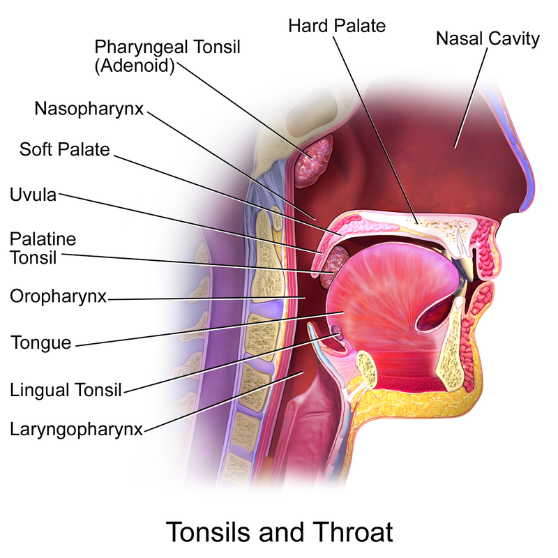
The palatine tonsils are the soft masses of lymphoid tissue located at the back of the throat that participate in both mucosal and systemic immunity [2]. The pharyngeal tonsils or adenoids are similar structures that can be found behind the nasal cavity. The adenoids and tonsils function as front line defenders against inhaled and ingested pathogens and the initial location for the generation of antigen-specific memory [3] [4]. Much like the appendix serves as a sentry for the colon, the tonsils serve as a sentry for the stomach, and with the appendix and tonsils both being components of the MALT (mucosa-associated lymphoid tissue), they are designed to work together [5] [6]. And tonsillar epithelial cells manufacture antimicrobial peptides termed defensins which play an important role in innate immune responses and help to shape the microbiota of the mouth [7]. Accordingly, drops in immunoglobulin levels after tonsillectomy may make vulnerable or predispose children to infectious diseases or allergic disorders, especially given that secretory IgA can be markedly reduced for more than 20 years after the tonsils are removed [8] [9] [10] [11]. We know that the tonsils play a large part in establishing and maintaining tolerance to allergens, and that deficiencies in vitamin A, vitamin D, and vitamin E are associated with the development of allergic disorders [12] [13] [14]. Specifically, vitamin D can increase the intratonsillar expression of the anti-inflammatory cytokine interleukin-37 (which helps modulate regulatory T cells and tolerance) as well as boost the production of the antiviral, antibacterial, and antifungal peptide LL-37 (also made by the tonsils) [15] [16] [17]. And vitamin A raises the tonsils’ crafting of interferon gamma, an immunomodulatory cytokine heavily involved in protection against viruses, bacteria, and protozoa [18]. The tonsils see their greatest immunological activity between the ages of 3 and 10, but childhood tonsillectomy is not without long-term consequence, as many conventionally-trained physicians still believe [19] [20]. In offering substantial examples as to why, a massive cohort study of almost 1.2 million children (published last year in the journal JAMA Otolaryngology – Head & Neck Surgery) revealed that having a tonsillectomy or adenoidectomy performed significantly heightened one’s risk for a range of respiratory, allergic, and infectious diseases (including asthma, pneumonia, influenza, chronic obstructive pulmonary disease, eczema, and urticaria or hives) [21]. Having the tonsils removed has also been associated with an increased risk of breast cancer and lymphoma, premature acute myocardial infarction (heart attack), Crohn’s disease, and appendicitis [22] [23] [24] [25] [26]. Rare complications of the tonsillectomy procedure itself include mediastinitis (inflammation of the mediastinum region between the lungs), Eagle syndrome (recurrent pain in the head and neck), atlantoaxial subluxation (partial dislocation among the first and second cervical vertebrae), and cervical osteomyelitis (infection of bone tissue in the cervical spine) [27]. Note that recurrent tonsillitis can be driven by upper cervical misalignment, so if this issue is present obviously it should be corrected before the tonsils are removed simply out of the diagnosis of recurrent tonsillitis [28]. Furthermore, let’s think for a minute – if the tonsils are repeatedly becoming inflamed, is it because they are dysfunctional, or is it because they are overwhelmed? If the tonsils are overwhelmed, will excising them eradicate the global burden on the immune system which overwhelmed them? Lastly, it is well known that between 1955 and 1963 both the oral and inactivated poliovirus vaccines were contaminated with simian virus 40 (SV40), a highly oncogenic DNA virus naturally hosted by rhesus monkeys that can induce the formation of tumors in brain, bone, lymph, and mesothelial tissue in humans [29] [30]. During the above time period, roughly 90% of children and 60% of adults in the United States were administered a poliovirus vaccine, and increased rates of multiple types of cancer have been found among those who were exposed to the contaminated vaccines [31] [32]. Most relevant to our interests is the fact that very clear evidence from research conducted decades ago (before the release of the Salk polio vaccine in 1955) demonstrated a significantly greater occurrence of bulbar poliomyelitis in those who had their tonsils removed versus those who had not [33] [34] [35]. In conclusion, I have not stated that the tonsils should never be removed. But the two main indications for tonsillectomy, recurrent throat infection and sleep-disordered breathing, are symptoms of deeper problems, and these deeper problems are not appropriately and completely remedied through focusing on the amelioration of enlarged tonsils. The tonsils and adenoids are laundering organs, so when morbid matter accumulates in the body, they can be called upon to assist in the elimination of refuse. Ergo, an overwhelming of the tonsils and adenoids will result in their congestion and inflammation. If congested and inflamed tonsils are extracted by an ENT, the body’s armament of laundering organs will be weakened, and remaining noxious material may find its way into the lungs or the gut. And this is why we find elevated incidences of respiratory and gastrointestinal ailments such as Crohn’s disease, appendicitis, asthma, pneumonia, and chronic obstructive pulmonary disease in those who have undergone a tonsillectomy. In vain does Nature protest against local symptomatic treatment, so please just be aware that a holistic and natural approach to improving the health of the body can prevent the need for a child’s tonsils to be taken out, despite the orthodoxy’s lack of understanding. References:
|
AuthorDenton Coleman is an Exercise Physiologist and Medical Researcher. Archives
October 2023
Categories |

 RSS Feed
