 Per Drs. Rod Jackson, Joanna Broad, Jennie Connor, and Susan Wells of the University of Auckland, “Do not assume there is a window in which the health benefits of alcohol are greater than the harms…” [1].
It is common to hear conventionally-trained cardiologists and other healthcare providers recommend a reasonable intake of wine or state that a periodic consumption of alcohol can actually help prevent against the onset of cardiovascular disease. Unfortunately, the notion of alcohol being beneficial for the heart is misguided and dangerous, and in this short article I’ll explain why. Firstly, the majority of evidence that seemingly supports a low level of alcohol being in the diet comes from epidemiological or observational studies, and as Jayasekara et al. have stated, “The quality of the epidemiologic evidence relating to a protective effect of low-dose alcohol consumption has been challenged” [2]. Much of the impact of alcohol drinking on mortality and the progression of disease states has been misinterpreted or viewed narrow-mindedly, as a large meta-analysis published in 2014 has explained: “These findings suggest that reductions of alcohol consumption, even for light to moderate drinkers, may be beneficial for cardiovascular health. Our results therefore challenge the concept of a cardioprotective effect associated with light to moderate alcohol consumption reported in observational studies and suggest that this effect may have been due to residual confounding or selection bias” [3]. Uncontrolled confounding can account for at least some, if not all, of the reported cardioprotective associations in non-randomized studies, as concluded in a paper by Naimi et al., “These findings suggest that some or all of the apparent protective effect of moderate alcohol consumption on CVD may be due to residual or unmeasured confounding” [4]. The results of observational studies can also be distorted by underlying poor health in nondrinkers and the existence of greater underlying health in light and moderate drinkers versus heavy drinkers. Further corroboration has been offered by Shaper, Wannamethee, and Walker: “The data suggest that the observed alcohol-mortality relationships are produced by pre-existing disease and by the movement of men with such disease into non-drinking or occasional-drinking categories. The concept of a “protective” effect of drinking on mortality, ignoring the dynamic relationship between ill-health and drinking behaviour, is likely to be ill founded” [5]. We must also refrain from looking at variables in isolation, for the effect of alcohol on HDL (high-density lipoprotein) cholesterol and platelet aggregation can only be considered positive if we ignore all of alcohol’s negative effects on the cardiovascular system and the body at large [6]. In that vein, let’s now examine the physiological consequences of tippling. Moderate alcohol intake can increase plasma triglyceride concentration which can heighten cardiovascular disease risk in men and women independent of HDL cholesterol changes [7] [8]. Normally, plasma triglycerides are hydrolyzed by the enzyme lipoprotein lipase in capillary endothelial cells, but alcohol can inhibit this enzyme and keep plasma triglycerides elevated by hindering the typical maturation of VLDLs (very-low-density lipoproteins) to IDLs (intermediate-density lipoproteins) and then to LDLs (low-density lipoproteins) as triglycerides are removed by lipoprotein lipase [9]. Thus, the consumption of alcohol can boost the plasma’s concentration of triglyceride-rich lipoproteins which can be atherogenic (TGRLs can worsen arterial wall permeability and cause vascular injury) [10] [11]. Alcohol can also inhibit the secretion of VLDLs from the liver (promoting the development of fatty liver) by impairing hepatocyte microtubule formation or obstructing the synthesis of phosphatidylcholine [12]. Ideally, the secretion of VLDLs by the liver is adequate to thwart fatty liver disease but insufficient to cause an abnormal accumulation of triglycerides in the blood by way of overwhelming the circulatory system’s metabolizing of these lipid carriers. As a far better alternative to liquor, daily supplementation with niacin (vitamin B3) can greatly and safely drop plasma triglycerides and LDLs as well as raise HDLs [13]. In fact, niacin, as expressed by Carlson, “lowers the levels of all atherogenic lipoproteins – VLDL and LDL with subclasses as well as Lp(a) – and in addition it raises more than any other drug the levels of the protective HDL lipoproteins” [14]. Overarchingly, alcohol interferes with the normal metabolism of lipids in the body, and through multiple pathways this can encourage the development of cardiovascular disease [15]. Moving on to a comparison between grapes and wine, whole juice from purple grapes has been shown to decrease platelet aggregation, boost platelet-derived nitric oxide release, and lower superoxide (a strong free radical) production in vivo [16]. And red grape juice has been found to more effectively curb atherosclerosis and improve lipid and antioxidant parameters versus red wine or dealcoholized red wine [17]. Accordingly, we can attribute the genuine benefit to heart health from wine to the polyphenols found in grapes, and not to ethanol (ethanol can be directly toxic to the cardiovascular system and can raise blood pressure) [18]. Furthermore, resveratrol consumption through grapes but not through wine has been associated with a reduced breast cancer risk, while a heightened breast cancer risk has been associated with consuming red wine [19] [20]. Even a light intake of alcohol (less than or equal to one drink per day) can notably escalate oral cancer risk [21]. Furthermore, persistent wine intake can lead to dental erosion, and there is concern over the amount of toxic metals (like lead, cadmium, and arsenic) that can be found in various wines [22] [23]. Now there is no doubt that resveratrol itself can be greatly cardioprotective and beneficial to the body. Though it’s important to remember that resveratrol certainly is not the only acting polyphenol in wine and grapes [24]. Resveratrol can excellently exert anti-inflammatory, antioxidant, and vasodilation effects, in addition to being able to suppress tumor growth and protect neurons in the brain against damage [25] [26] [27]. More specifically, resveratrol helps to regulate redox homeostasis in humans and can block the oxidation of LDLs (drinking wine or other alcoholic beverages can up LDL oxidation), as well as induce apoptosis in cancer cells and better both insulin sensitivity and blood pressure [28] [29] [30] [31] [32]. After the ingestion of an alcoholic beverage, most of the ethanol will be oxidized in the liver by the enzyme alcohol dehydrogenase, yielding the highly toxic metabolite acetaldehyde [33]. Ideally, yielded acetaldehyde will be quickly converted into acetate by another liver enzyme, acetaldehyde dehydrogenase, with the help of glutathione. Acetate should then be converted into acetyl coenzyme A and ultimately into water and carbon dioxide. With a deficient supply of glutathione however, acetaldehyde can accumulate in the body and wreak havoc as it is a major generator of free radicals and a known carcinogen [34]. Also note that acetaldehyde occurs naturally in alcoholic beverages in addition to arising from ethanol metabolism [35]. We know that acetaldehyde directly impairs the contractile function of the heart, disrupts cardiac excitation-contraction coupling, and fosters lipid peroxidation and oxidative damage in the heart [36]. And ethanol metabolites termed ‘fatty acid ethyl esters’ can encourage injury to the liver, heart, and pancreas [37]. Acetaldehyde can also induce mitochondrial dysfunction and stress endoplasmic reticula, as well as negatively modify and form adducts with various proteins that stimulates the crafting of antibodies against acetaldehyde epitopes (which can lead to autoimmune responses) [38] [39] [40]. Acetaldehyde may contribute to organ damage (including the brain) by way of inflammatory cytokines and the same forming of protein adducts just mentioned too [41]. As a side note, acetaldehyde-induced dysfunction of the heart can be somewhat ameliorated by thiamine (vitamin B1) and folate (vitamin B9) [42]. Wrapping up my argument with a few more consequences of tippling, alcoholic beverages can suppress impeding wave motility in the small intestine (cutting the amount of time ingested food has to be properly digested), injure the gut mucosa and intestinal microbiota (potentially causing small intestinal bacterial overgrowth), magnify the translocation of bacterial toxins (like lipopolysaccharide) from the gut to the blood (which can contribute to liver and heart disease progression), and increase the permeability of the intestinal wall (creating a leaky gut) [43] [44] [45] [46] [47]. Importantly, ethanol (a carcinogen itself) can also facilitate the activation of procarcinogens (turning them into carcinogens) and the conversion of xenobiotics to toxic metabolites, enhancing the harmfulness of dietary and environmental toxins [48] [49]. Lastly, ethanol exposure can lead to atrophy of all skeletal muscle fiber types, lowered muscle capillarity, and altered metabolism in muscle tissue too [50]. In conclusion, I am not stating that alcohol, especially red wine, is the worst thing we could possibly put into our bodies. If you’d like to indulge in a drink here and there, by all means do your thing. Just please don’t assume that by knocking a few back you’re doing something to improve the health of your heart as some who should know better have suggested. Alternatively, simply eating some grapes and berries (plus some niacin-rich foods like chicken, turkey, sunflower seeds, green peas, and avocados) will beautifully supply your body with plenty to support your cardiovascular system and more without any of the ill effects of ethanol and its metabolites. Appropriately supplementing with isolated niacin and resveratrol would be another option for safely profiting the heart. Chalk another one up for food as medicine. References:
0 Comments
 For ages the salamanders have been considered a class of the elementals – vehicles of the transmuting fire element which they guard and embody. Dwelling in their subtle ether, these transubstantial nature spirits were often invoked and honored via the burning of incense. Later being associated with the element nitrogen as understood within modern chemistry, the salamanders are said to be operant in the emotions of man through the liver and bloodstream (the liver is believed to be the seat of the emotional principle and assists in processing emotions in addition to its biochemical transformation roles) [1]. Salamanders (as elementals) have been assigned the cardinal direction of south, and it is maintained that they are the most powerful of the nature spirits.
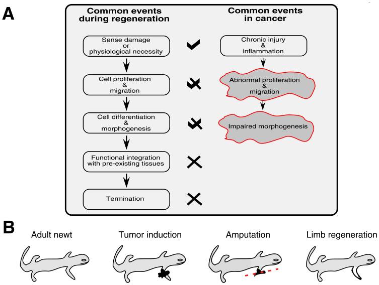
Salamanders are quite unique among vertebrates in their capacity to resist the development of cancer and regenerate limbs, and for many decades the link between regenerative ability and cancer protection has been discussed [2]. Normal tissue regeneration involves the directing of progenitor cells to an area of need, the careful proliferation and differentiation of those cells, and timely termination signals [3]. Aberrant or unregulated restoration efforts would obviously create unwanted consequences, but appropriate regeneration can actually halt and reverse the autonomous growth of malignant cells [4] [5] [6]. Because of the corollaries that exist between the events of tissue regeneration and the progression of cancerous growths, local tumors can be described as wounds that do not heal [7]. Chronic tissue stress or injury can drive ongoing inflammation which leads to an incomplete repair and restore operation that involves the misappropriating of mechanisms governing tissue mending and stem cell self-renewal – and this subversion fosters tumor maturation [8]. Because there are intracellular and extracellular factors that can regulate the self-renewal of stem cells, remedying the cancer pathology must include a reestablishment of normalcy and health to the microenvironment or terrain of the malignancy [9]. The remainder of this article will revolve around this concept as we look deeper into cancer’s idiosyncrasies. The tumor-suppressing protein p53 is frequently inactivated by mutations or viral oncogenes in human cancers, and Villiard et al. have shown that p53 signaling is necessary for limb regeneration in salamanders [10] [11]. And a related study provided evidence that more robust p53 proteins and a greater number of them confers heightened tumor resistance [12]. Ordinarily, p53 activation triggers DNA repair, apoptosis (mainly through the mitochondrial death pathway), and senescence signaling which helps to eliminate faulty cells and thwart cancerous growths [13]. Additionally, the p53 protein normally checks the self-renewal of adult stem cells, so when this protein is dysfunctional or inactive, stem cells will have less of a controlling leash, and therefore may abnormally proliferate more easily [14]. Again, the events of regeneration and tumorigenesis can be seen as two different but related paths. When the p53 protein is functioning as it should, it blocks the initial steps of regeneration from shifting toward tumorigenesis [15]. Thus, correct p53 activity helps prevent the transforming of healthy tissue restoration into unhealthy tissue malignancy. Moving forward, well-orchestrated sequences are necessary for the molding of replacement tissue in adult vertebrates to prohibit errant growth of mature cells that have dedifferentiated to fuel healing efforts. Salamanders skillfully employ dedifferentiation and transdifferentiation of mature cells to rebuild tissues that have been damaged or removed [16]. As Oviedo and Beane have explained, “in mammals chronic epithelial injury often precedes malignant transformation, while in urodeles (salamanders) and planarians (flatworms) persistent damage generally ends merely with functional repair. Injury response always entails attempts to repair the damage, but the fundamental differences probably lay in the coordination of such responses rather than in structural or species-specific capabilities” [17]. And evidence suggests that it is “individuation” or regenerative fields that direct and coordinate epimorphic regeneration in animals – a notion put forth by Conrad Hal Waddington in 1935 [18]. In 1938, Burr, Smith, and Strong found that bioelectric differences exist between cancer-susceptible and cancer-resistant mice, as well as that the onset of cancer is associated with a global deviation in an organism’s electrodynamic field [19]. Accordingly, we can state that cancer’s pathology is not merely the result of a random somatic mutation, and that it indeed consists of informational as well as molecular elements. Continuing, remodeling of the extracellular matrix is a major piece of limb regeneration and as a side note this process can be augmented with sufficient vitamin A [20]. Fibroblasts are prototypical cells that construct extracellular matrices and play central roles in both wound healing and tumor formation [21]. In salamanders, the dedifferentiation of fibroblasts (as well as some other cell types) after injury gives rise to what is known as a regeneration blastema (a mass of undifferentiated cells primed to repair lost tissue) that supplies the injured area with restorative cells [22]. In humans, the wound healing process can be divided into four phases: hemostasis, inflammation, proliferation, and tissue remodeling [23]. As the proliferation phase concludes in humans, certain fibroblasts (myofibroblasts) undergo apoptosis and are then cleared by macrophages [24]. In cancer, instead of being cleared away by macrophages, myofibroblasts remain in the diseased or injured region and become cancer-associated fibroblasts that are persistently activated by and promote the expansion of the developing tumor [25]. Now, earlier I mentioned the importance of reestablishing normalcy and health to the microenvironment of a tumor in the reversing of cancer because communication between neoplastic cells and their microenvironment drives tumor progression [26]. For corroboration, Bissell and Hines have explained that “the microenvironment can provide crucial signaling to maintain tissue architecture, inhibit cell growth and suppress or revert the malignant phenotype…incorrect signals from the microenvironment should lead to destabilization of tissue homeostasis and initiation and promotion of normal cells to malignancy” [27]. Salamanders masterfully create embryonic-like microenvironments at the site of regeneration after injury, and this not only ensures the correct reconstructing of harmed or missing tissue, but can also reprogram cancer cells (via signaling pathways) so that they revert to a normal phenotype [28]. Such reprogramming can also take place in the human body, and the homeobox genes (which code for proteins that help direct morphogenesis) that salamanders utilize in regeneration are also present in humans [29] [30]. Appropriate activation of beta-catenin and wnt signaling could trigger expression of the above genes that leads to commands and instructions for regeneration being given in Homo sapiens [31]. Alternatively, virus-mediated transduction could be used to extract functional homeobox genes from salamanders and insert them into human cells at sites requiring healing [32]. Immunomodulatory mesenchymal stem cells can be found throughout the body, and correct regeneration of human fingertips has been seen, in addition to the regenerative capacities displayed by human endometrium, kidney, liver, bladder, lung, gut, bone, skeletal muscle, and heart tissue [33] [34] [35]. So the commonly propagated idea that we uprights are largely incapable of true regeneration is incorrect. Digging deeper still, nerve dependence refers to the outgrowth of axons being necessary for the reconstituting of amputated limbs in animals [36]. In order for epimorphic regeneration to proceed rightly in some organisms, there must be a presence of regenerating nerves in the progenitor cell niche or microenvironment [37]. Interestingly, denervation of a neoplasm can stunt its expansion and suppress metastasis, suggesting nerve dependence in cancer [38]. Once more, there are parallels between the steps of regenerative healing and cancer, and it appears that the interaction of sprouting nerves with blastema and tumor cells is similar, meaning the innervation of proliferating tissue is capable of promoting either regeneration or tumorigenesis depending on the conditions of and signaling within the local microenvironment [39]. Evidently, neural facilitation of cancer progression includes the release of trophic factors by nerves toward tumor and stroma cells, the release of trophic factors by tumor cells beckoning nerve infiltration, activation of the stem cell compartment by neurotransmitters, and the supporting of neoangiogenesis [40]. So, in both regenerative blastemas and malignancies, there is bidirectional communication between sprouting nerves and proliferating cells [41]. Extrapolating a bit, we know that mitochondria can be transferred between cells in mammals, and that sabotaged mitochondrial respiration can be restored in neoplastic cells via the acquisition of mitochondria from other cells or the local microenvironment (which propagates tumor advancement) [42] [43]. Because of the known interaction between nerves, tumor cells, and microenvironments, and because nerve fibers reach into virtually every crevice of the body, the nervous system may not only aid in the apportioning of mitochondria to target tissues but also stand as an overseer in both regeneration and cancer. Coming to the last stop on our train ride, it is clear that mitochondrial dysfunction from free radicals or toxins, harmful electromagnetic radiation, hypoxia, viral infections, or abnormal mtDNA gene expression can induce a kind of metabolic reprogramming that mirrors the Warburg effect of cancer cells relying on glycolysis for their making of energy instead of oxidative phosphorylation and includes the acquisition of an invasive phenotype [44]. The arising of defects in mitochondria then may kickstart a signaling cascade that fosters tumor formation, whereas healthy mitochondria can suppress tumorigenesis [45]. As Seyfried et al. have expressed, “Any unspecific condition that damages a cell’s respiratory capacity but is not severe enough to kill the cell can potentially initiate the path to a malignant cancer” [46]. So the ability of cells to properly breathe is very important for a state of hypoxia or low tissue oxygen can initiate cell death via apoptosis (with the help of functional p53 proteins) or provoke adaptive responses that contribute to tumor formation [47]. And a hypoxic state encourages lactate production via glycolysis and subsequent tissue acidosis, which favors neoplasm progression and obstructs antitumor immunity (hypoxic and acidic extracellular spaces also propel invasion and metastasis) [48] [49]. Note that both chemotherapy and radiation therapy can damage mitochondrial respiration in healthy cells, negatively alter the tumor microenvironment, and encourage cancer recurrence [50] [51] [52] [53] [54] [55] [56]. Significant evidence supports the hypothesis that essentially all cancer hallmarks can be linked to impaired mitochondrial function and energy crafting [57]. In other words, cancer should be viewed primarily as a metabolic disease and not a genetic disease. Finally, the integrity of the nuclear genome leans heavily upon efficient and normal mitochondrial function as mitochondrial stress can engender DNA repair abnormalities and the upregulation of ATP making via glycolysis [58] [59] [60]. And this brings us full circle because the metabolic yields of high-rate glycolysis can destabilize the morphogenetic field of a microenvironment and contribute to inflammation and tumor advancement [61] [62]. We can equate ‘morphogenetic field’ to the term individuation field introduced earlier, and therefore conclude that individuation fields coordinate epimorphic regeneration when stable and healthy, yet advocate the manifestation of cancer when unstable and infirmed because of metabolic ill-health (resulting from such factors as hypoxia, acidosis, nutrient deficiencies, toxicity, and inflammation) with mitochondria and ATP production as the gateway. ATP is the energy of life, so when an organism becomes incapable of effectively and efficiently crafting ATP, the stage can be set for cancer’s appearance. Just as the salamander remains aligned with and ensconced in life by transmuting that which obstructs life, so too may we unhinder our ability to heal and forestall cancer by augmenting our transmutative faculty through eating alive foods, drinking alive water, shielding ourselves from discordant EMFs, detoxifying regularly, and staying positive in thought and feeling. The appropriate prevention and treatment of cancer then, lies not in the worsening of the body’s health and its power to make ATP, but in the improving of the body’s health and its power to make ATP – and this is why natural oncology experiences the success it does. References:
 In the first half of the previous century, tonsillectomy was the most frequently performed surgical procedure in America, and is still one of the most common today [1]. Yet surgical excision of the tonsils and/or adenoids is not as harmless or valid as commonly thought, and in this short article we’ll examine why.
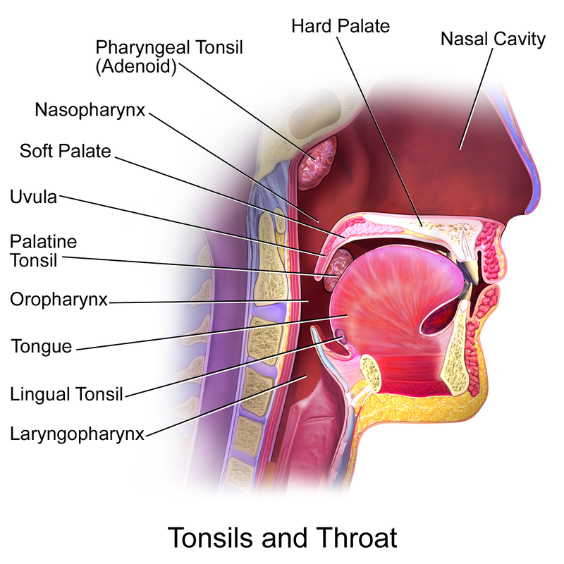
The palatine tonsils are the soft masses of lymphoid tissue located at the back of the throat that participate in both mucosal and systemic immunity [2]. The pharyngeal tonsils or adenoids are similar structures that can be found behind the nasal cavity. The adenoids and tonsils function as front line defenders against inhaled and ingested pathogens and the initial location for the generation of antigen-specific memory [3] [4]. Much like the appendix serves as a sentry for the colon, the tonsils serve as a sentry for the stomach, and with the appendix and tonsils both being components of the MALT (mucosa-associated lymphoid tissue), they are designed to work together [5] [6]. And tonsillar epithelial cells manufacture antimicrobial peptides termed defensins which play an important role in innate immune responses and help to shape the microbiota of the mouth [7]. Accordingly, drops in immunoglobulin levels after tonsillectomy may make vulnerable or predispose children to infectious diseases or allergic disorders, especially given that secretory IgA can be markedly reduced for more than 20 years after the tonsils are removed [8] [9] [10] [11]. We know that the tonsils play a large part in establishing and maintaining tolerance to allergens, and that deficiencies in vitamin A, vitamin D, and vitamin E are associated with the development of allergic disorders [12] [13] [14]. Specifically, vitamin D can increase the intratonsillar expression of the anti-inflammatory cytokine interleukin-37 (which helps modulate regulatory T cells and tolerance) as well as boost the production of the antiviral, antibacterial, and antifungal peptide LL-37 (also made by the tonsils) [15] [16] [17]. And vitamin A raises the tonsils’ crafting of interferon gamma, an immunomodulatory cytokine heavily involved in protection against viruses, bacteria, and protozoa [18]. The tonsils see their greatest immunological activity between the ages of 3 and 10, but childhood tonsillectomy is not without long-term consequence, as many conventionally-trained physicians still believe [19] [20]. In offering substantial examples as to why, a massive cohort study of almost 1.2 million children (published last year in the journal JAMA Otolaryngology – Head & Neck Surgery) revealed that having a tonsillectomy or adenoidectomy performed significantly heightened one’s risk for a range of respiratory, allergic, and infectious diseases (including asthma, pneumonia, influenza, chronic obstructive pulmonary disease, eczema, and urticaria or hives) [21]. Having the tonsils removed has also been associated with an increased risk of breast cancer and lymphoma, premature acute myocardial infarction (heart attack), Crohn’s disease, and appendicitis [22] [23] [24] [25] [26]. Rare complications of the tonsillectomy procedure itself include mediastinitis (inflammation of the mediastinum region between the lungs), Eagle syndrome (recurrent pain in the head and neck), atlantoaxial subluxation (partial dislocation among the first and second cervical vertebrae), and cervical osteomyelitis (infection of bone tissue in the cervical spine) [27]. Note that recurrent tonsillitis can be driven by upper cervical misalignment, so if this issue is present obviously it should be corrected before the tonsils are removed simply out of the diagnosis of recurrent tonsillitis [28]. Furthermore, let’s think for a minute – if the tonsils are repeatedly becoming inflamed, is it because they are dysfunctional, or is it because they are overwhelmed? If the tonsils are overwhelmed, will excising them eradicate the global burden on the immune system which overwhelmed them? Lastly, it is well known that between 1955 and 1963 both the oral and inactivated poliovirus vaccines were contaminated with simian virus 40 (SV40), a highly oncogenic DNA virus naturally hosted by rhesus monkeys that can induce the formation of tumors in brain, bone, lymph, and mesothelial tissue in humans [29] [30]. During the above time period, roughly 90% of children and 60% of adults in the United States were administered a poliovirus vaccine, and increased rates of multiple types of cancer have been found among those who were exposed to the contaminated vaccines [31] [32]. Most relevant to our interests is the fact that very clear evidence from research conducted decades ago (before the release of the Salk polio vaccine in 1955) demonstrated a significantly greater occurrence of bulbar poliomyelitis in those who had their tonsils removed versus those who had not [33] [34] [35]. In conclusion, I have not stated that the tonsils should never be removed. But the two main indications for tonsillectomy, recurrent throat infection and sleep-disordered breathing, are symptoms of deeper problems, and these deeper problems are not appropriately and completely remedied through focusing on the amelioration of enlarged tonsils. The tonsils and adenoids are laundering organs, so when morbid matter accumulates in the body, they can be called upon to assist in the elimination of refuse. Ergo, an overwhelming of the tonsils and adenoids will result in their congestion and inflammation. If congested and inflamed tonsils are extracted by an ENT, the body’s armament of laundering organs will be weakened, and remaining noxious material may find its way into the lungs or the gut. And this is why we find elevated incidences of respiratory and gastrointestinal ailments such as Crohn’s disease, appendicitis, asthma, pneumonia, and chronic obstructive pulmonary disease in those who have undergone a tonsillectomy. In vain does Nature protest against local symptomatic treatment, so please just be aware that a holistic and natural approach to improving the health of the body can prevent the need for a child’s tonsils to be taken out, despite the orthodoxy’s lack of understanding. References:
 Forest bathing, or shinrin-yoku in Japanese, is a term coined by Tomohide Akiyama for the simple practice of connecting to and taking in nature through most if not all of the physical senses [1]. By reengaging with nature we reattune ourselves with its rhythms and reinvigorate our bodies by stepping back into harmony. As Dr. Qing Li has spoken to, universal scripture is written in nature for the holy book of God is the natural world itself [2].
Spending time in a tranquil, outdoor setting allows for ‘involuntary attention’ or ‘soft fascination’ whereby the mind may rest and our capacity for clear cognition may be restored [3]. Forest medicine kindles our faculty of self-healing, and in 1984, Dr. Roger Ulrich published a paper in the journal Science which reported that patients assigned to hospital rooms with windows looking out on a natural scene experienced enhanced recovery from surgery (multiple subsequent studies confirmed his findings) [4]. As the work of Dr. Richard Taylor has shown, viewing of the fractal patterns ubiquitous in nature can reduce physiological stress and beneficially alter brain wave activity [5] [6]. Being a student of Plato, Aristotle surely understood the health-giving and divine quality of the living world when he stated that “In all things of nature there is something of the marvelous.” Through forest bathing we gift ourselves with the original means of aromatherapy and inhale an abundance of phytoncides from surrounding trees and plants [7]. Phytoncides are volatile compounds that can have antimicrobial, antioxidant, and anti-inflammatory effects and which play vital roles in air purification and communication within ecosystems [8] [9]. The air around rivers, streams, and woodlands is also rich with negatively-charged ions, a healthy uptake of which can protect the body against stress exposure, exert an anti-cancer effect, and enhance activity of the antioxidant enzyme superoxide dismutase [10] [11] [12]. Forest bathing also brings us into contact with the healthy microbes we have coevolved with, one example of such being Mycobacterium vaccae. Mycobacterium vaccae is a nonpathogenic mycobacterium species found normally in healthy soil that can serve as a beneficial psychobiotic when ingested or inhaled [13]. Mycobacterium vaccae can notably impact the gut-brain axis and bring about a decrease in anxiety, an increase in learning efficacy, and an improvement in immune responses (mycobacteria have been studied as immunotherapy agents) [14] [15]. Furthermore, M. vaccae has shown promise in the treatment of allergic disorders, and it can be argued that a lack of being exposed to this mycobacterium from our modern way of living is partly responsible for the heightened prevalence of allergic disorders we now see [16] [17] [18]. Through various means, the practice of shinrin-yoku is also capable of lowering both blood pressure and blood glucose, bettering depression and mental health, boosting the number of natural killer cells and the making of intracellular anti-cancer proteins, raising parasympathetic tone and lowering cortisol, dropping LDL cholesterol, heightening working memory performance, and simply improving overall well-being [19] [20] [21] [22] [23] [24] [25] [26] [27] [28]. In conclusion, connecting with nature irrefutably profits the mind, body, and spirit, and there is no doubt that forest medicine will continue to grow as a beautiful bestowal in both healthcare and transcendentality [29]. References:
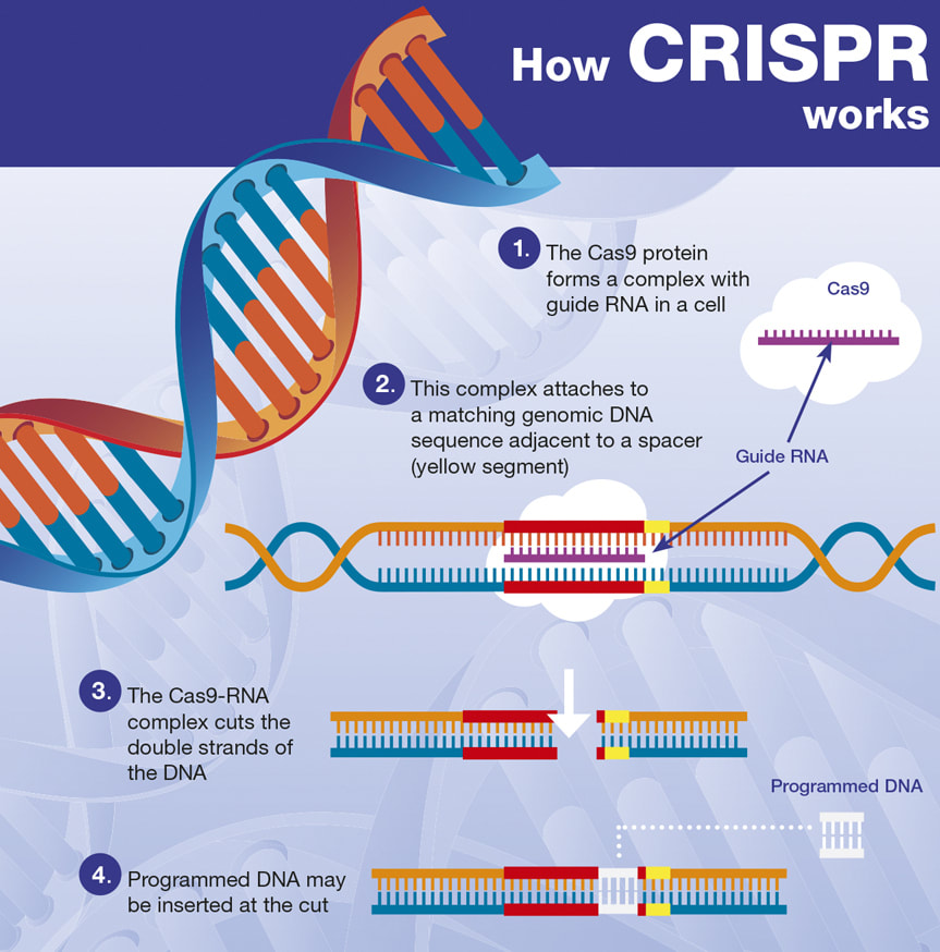 CRISPR is an acronym for ‘clustered regularly interspaced short palindromic repeats,’ which are nucleotide sequences and spacers found in specialized regions of DNA from prokaryotic organisms (usually single-celled bacteria or archaea that lack a membrane-bound nucleus and membrane-bound organelles) [1]. These regions are accompanied by Cas proteins and together they make up antiviral utilities which detect and degrade exogenous DNA and RNA from infiltrating viruses (there is also evidence of CRISPR-Cas systems helping with endogenous transcriptional control) [2]. CRISPR sequences originate in prokaryotes from past viral infections or plasmid exposures, and Cas (CRISPR-associated) proteins are enzymes that use CRISPR sequences to specifically locate and cleave matching or complimentary DNA fragments to thwart subsequent infection or unwanted genome alterations through the uptake of foreign nucleic acids [3] [4].
The CRISPR-Cas system can be hijacked or differently employed as a gene therapy tool, and this fact has led to the development of a gene editing technology named CRISPR-Cas9 (in which one particular Cas protein, Cas9, is utilized) [5]. CRISPR-Cas9 allows for targeted DNA deletions, insertions, and substitutions, and thus can be compared to a word processor’s cut and paste function. Viral means and non-viral means (like lipid- or polymer-based nanocarriers) for delivering the CRISPR-Cas9 vehicle to target cells currently exist (note that viral vectors carry a risk of cancer induction and violent immune responses) [6]. And a related technology involving nuclease-deactivated Cas9 (CRISPR-dCas9), can be used to epigenetically alter the activity of a particular gene without the normal DNA-cutting action of Cas9 [7]. CRISPR-Cas9 is not a perfect instrument however, as there can be variable accuracy and efficiency issues. And because gene editing literally consists of making changes to an organism’s genetic code, there are major ethical concerns with its extrapolated use (beyond simply correcting genetic defects in those with an inherited genetic disease and into extensions like germline engineering) [8]. Now, Duchenne muscular dystrophy (DMD) stands as an inherited genetic disease in which mutations in the DMD gene typically cause a failure to manufacture the protein dystrophin, an integral component of the dystrophin-glycoprotein complex found in heart and skeletal muscle cells that serves as a cytoskeleton-extracellular matrix bridge [9]. Without the mechanical stabilization and signaling roles of the dystrophin-glycoprotein complex, cardiac and skeletal myocytes degenerate with use and progressive muscle wasting results [10]. DMD patients typically experience cardiac or respiratory failure during the third or fourth decade of life resulting in premature death [11]. The correction of DMD mutations via exon skipping or exon deletion with the use of CRISPR-Cas9 can restore expression of the dystrophin protein and rescue the function of cardiac and skeletal muscle cells, and this has been safely demonstrated in mouse models of Duchenne muscular dystrophy [12] [13] [14]. Other strategies for remedying DMD mutations with the help of CRISPR-Cas9 are available too [15]. DMD mutations have also been corrected using CRISPR-Cas9 in human cells in vitro [16] [17] [18] [19] [20]. And dystrophin expression and improved muscle histology have been seen in a canine model of DMD [21]. While thousands of DMD mutations have been identified in patients with Duchenne muscular dystrophy, CRISPR technologies have exhibited efficacy in restoring dystrophin expression in human cells, and thus offer great promise for an otherwise incurable disease [22]. In conclusion, even though CRISPR systems remain highly advantageous because they are capable of permanently fixing genetic defects in those with DMD, there is an unpredictability to gene editing, and undesired mutations, off-target effects, and dangerous immune responses can be seen [23]. Genetic mosaicism has helped teach us that the human genome is much more malleable than previously believed, which carries with it both good and bad potentials [24]. Gene editing has the capacity to powerfully treat and even rectify many inherited genetic diseases, but certainly we must be careful to not get lured into playing God. Kept within strict boundaries, gene therapy can continue to grow into a medical revolution, and very soon, CRISPR systems may become easily accessible to those who truly need them. References:
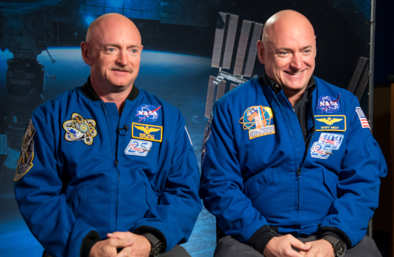 Embarking in 2015, NASA astronaut Scott Kelly spent one year aboard the International Space Station while his twin brother Mark remained on Earth. Blood, saliva, urine, and stool samples were collected from both Scott and Mark during the one-year mission. While a variety of physiological markers and assessments were analyzed, two discrepancies are relevant to our interests. Firstly, a handful of Scott’s genes experienced a change in their expression during his time in space that failed to return to baseline after six months of arriving back on Earth [1]. Such an alteration is to be expected from spending a year in the stressful conditions of near-Earth space, but we will draw important implications from this finding. Secondly, the average length of Scott’s telomeres increased significantly aboard the International Space Station, an unexpected finding given that these chromosomal endcaps normally shorten with age due to a snipping that takes place with repeated cell division (though it is possible to lengthen telomeres with diet, exercise, and stress management) [2]. Upon returning to Earth, Scott’s average telomere length quickly stabilized to preflight levels.
So how do these discoveries relate to spirituality? A super quick dive into biophysics will reveal the answer. It is well known that extended exposure to a microgravity environment from spaceflight can suppress or hamper the human immune system by impacting cytoskeletons [3]. As Littleton and Ludwick suggest, cytoskeletons function as cellular gravity sensors, so microgravity exposure can muddle the regulation performed by cytoskeletons and disrupt cell motility, cell signaling, cell proliferation, and gene expression [4]. Mechanical stress or distortion stimuli are converted into chemical signals (a process termed mechanotransduction) by proteins comprising cytoskeletons and extracellular matrices [5]. Because there are physical connections between all of the tissue systems in the body, physiological changes at the organism level can be seen during spaceflight or extended exposure to microgravity [6]. Now, we know that the assembly or self-organization of microtubules (polymeric cytoskeleton components) depends on the force of gravity [7]. We also know that information can be encoded within the assembly pattern of tubulin monomers inside microtubules [8]. Furthermore, as Ostovari, Alipour, and Mehdizadeh have proposed, quantum entanglement between tubulin monomers and emitted biophotons may allow for the transmittance and exchange of both stored and accessed information throughout the body [9]. According to Sorli et al., the evolution of life is encoded within higher-dimensional Hilbert spaces and conveyed to our third dimension via biophotons [10]. Because of the quantum entanglement between microtubules and biophotons, microtubules can also partake in nonlocal interactions and transmit higher-dimensional data to the physical body through the medium of Hilbert space “pilot photons” [11]. Such data may be shuttled from biophotons to chromosomes through the vehicle of microtubule-associated proteins – allowing for a direct change to one’s DNA by way of the mind [12]. Furthering the above, physical evidence exists for gravity being able to carry with it electromagnetic data [13] [14]. And this forces us to correct the conventional definition of gravity, which is not simply one of the four forces of nature which causes apples to fall on our heads when we sit underneath trees [15]. Gravity is the toward-unity movement of consciousness units, the inward spiraling of the aether wind as the Creator wraps its arms around the universe and pulls All That Is back to it. Gravity is love, the harmonization with oneness. In other words, gravity is one half of the breath of Brahma, the inbreathing and outbreathing of the cosmos. As the planet continues onward with its ascension, additional instreams will flow from the center of the Milky Way galaxy, pass through our sun, and be delivered to the Earth. These instreams will further our consciousness elevation and greatly assist in guiding us back home. In conclusion, now we can see that we are co-creating, spiritual beings who are currently participating in a physical reality that is shaped by gravity and therefore malleable to our openness to love. The universe is perfect, but when we separate ourselves from love we perceive imperfection because we are no longer looking through the eyes of God. The completing of Earth’s evolution will entail a return to love because there is no escaping the oneness of creation. To love yourself is to love God, and to see God in all things is to see the universe as it truly is. References:
 Iodine is an electronegative element of the halogen family, and is an essential nutrient that must be obtained through dietary sources. Iodide is the anionic or negatively-charged form of iodine. The iodine content of foods is largely dependent upon the iodine content of the soil in which they are grown. Most foods are naturally quite low in iodine, save for most saltwater fish and various sea vegetables or seaweed [1] [2]. Undoubtedly, iodine deficiency continues to stand as a significant health issue throughout the world, affecting both industrialized and developing nations [3]. The propagandization of iodized salt is objectionable though, as conventional table salt is crap, and iodized salt can lose much of its added iodine during storage [4] [5].
Because of the need for iodine in the making of thyroid hormones, iodine is necessary for the normal development of the early brain and for general growth and maturation (in addition to the maintenance of a healthy basal metabolic rate) [6]. Iodine deficiency is one of the most common causes of preventable cognitive impairment, with a moderate-to-severe lack of iodine being associated with a reduction in mean IQ scores of 13.5 points [7] [8]. And the classical marker of iodine deficiency is enlargement of the thyroid gland or goiter [9]. Sufficient iodine intake during pregnancy is crucial, for the requirement of this nutrient rises by about 50% during the gestation period [10]. Appropriate supplementation with or adequate intake of iodine can prevent cretinism, increase birth weight, and decrease infant mortality [11]. The recommended level for iodine intake during pregnancy is at least 250 micrograms per day [12]. Ingested iodide is efficiently absorbed in the stomach and small intestine [13]. Most ingested iodine is converted into iodide in the gut before being absorbed into the bloodstream, but not all [14] [15]. So supplementing with a combination of iodide and iodine can be more effective or beneficial than supplementing with iodide (usually in the form of potassium iodide) alone. Also, selenium and iron concentrations need to be adequate in order for iodine to be properly utilized in the thyroid gland [16]. Iodide absorbed into the bloodstream quickly mixes with iodine obtained from the breakdown of iodothyronines (T3 and T4) to form the extrathyroidal pool of ‘plasma inorganic iodine,’ regulated by the thyroid and kidneys [17]. Evidently, a normal adult employs roughly 80 micrograms of iodine per day in the manufacturing of the thyroid hormones, but much more than 80 micrograms of iodine is required by the body each day [18]. For adults, the recommended daily allowance (RDA) for iodine is 150 micrograms per day, but because of our exposure to iodine-blocking pollutants, it could be argued that such a level is too low for modern needs [19]. Furthermore, we should consider that many Japanese citizens consume far more than 150 micrograms of iodine per day, and that breast cancer incidence and mortality rates have historically been much lower in Japan compared to the United States [20] [21]. A 24-hour urinary iodine test can be used to assess iodine status, but because most ingested iodine is excreted through the urine within 24 hours, results from this test can vary quite a bit [22]. Hence, iodine sufficiency is tough to measure accurately without collecting multiple urine samples [23]. And it’s worth noting that athletes or anyone who participates in vigorous exercise can lose a notable amount of iodine through sweat, so iodine supplementation could be warranted in these individuals [24]. Iodide uptake in the thyroid gland and elsewhere is accomplished via sodium-iodide symporters (membrane proteins that traffic iodide into cells), which are located mostly in the thyroid, but can also be found in the stomach, small intestine, breasts, and salivary glands [25]. Normal sodium-iodide symporter (abbreviated as NIS) function can be disturbed as a result of inflammation, a genetic defect, or exposure to environmental toxins like perchlorates and phthalates [26] [27] [28] [29] [30]. Evidence suggests that vitamin C supplementation works well for helping to repair dysfunctional sodium-iodide symporters [31]. Despite claims to the contrary, the presence of excessive iodide in the body can suppress thyroid function by inhibiting the NIS-mediated uptake of iodide in the thyroid [32] [33]. This suppression of thyroid hormone synthesis should be temporary, but there can be a failure to “escape” from the Wolff-Chaikoff effect in which the suppression is prolonged or sustained (those with autoimmune thyroiditis are more susceptible to this failure) [34]. Too much iodine can stifle autophagy in the thyroid and promote apoptosis of thyroid follicular cells as well [35]. Iodide excess can also downregulate the NIS-mediated uptake of iodide in the small intestine [36]. With all of that said, a molecular iodine dosage of 6,000 micrograms per day for five months has been administered to healthy, euthyroid women without indication of toxicity [37]. Wagner et al. observed that the maximum accumulation of iodide by the thyroid gland was achieved at a dosage of 600 micrograms per day [38]. And it has been concluded by Koutras et al. that “as the plasma inorganic iodine level rises, iodide utilization by the thyroid becomes less complete” [39]. A relatively recent estimate of total body iodine content averaged 14.6 milligrams (not to be confused with micrograms, 1 milligram equals 1,000 micrograms), the majority of iodine being concentrated in the thyroid gland [40]. Now let’s turn our attention to the known roles of iodine in the body and potential benefits that might be derived from its supplementation. Firstly, it’s important to know that bromide can weaken the impact of consumed iodide by decreasing the accumulation of iodide in the thyroid gland and increasing the excretion of iodide through the kidneys (bromine can be found in some cosmetics as well as some plastics and flame retardants) [41]. And the potency of fluoride as an antagonist to iodine is well known, with fluoride’s deleterious effects on the thyroid gland and brain being strong enough to masquerade as serious iodine deficiency [42] [43]. Chlorine and organochlorine pesticides may antagonize iodine in the body similar to fluoride too [44] [45]. Fortunately, iodine supplementation can greatly assist in the displacement and flushing out of the halides bromide and fluoride [46]. Abraham has reported that the giving of an iodide and iodine combination supplement can also promote the excretion of the toxic metals lead, mercury, cadmium, and aluminum [47]. Secondly, acute damage to the mucosa of the esophagus and stomach has been documented with the application of Lugol’s iodine, but this occurrence is uncommon [48] [49]. Placing drops of a liquid iodine solution into an enteric-coated capsule might bypass this potential issue. Moving on, iodide can serve as an antioxidant by scavenging hydroxyl radicals and defending against lipid peroxidation in cell membranes [50] [51]. The thymus gland concentrates inorganic (nonradioactive) iodine, and iodine supplementation appears to strengthen the adaptive immune system [52] [53]. Iodine can also reduce the virulence of H. pylori bacteria in the stomach as well as help normalize corticosterone secretion throughout the day [54] [55]. Stable iodine can thwart the thyroid’s uptake of radioactive iodine-131 present in nuclear fallout, but it won’t protect the body against other radionuclides like cesium-137, strontium-90, and plutonium-241 [56]. Iodine supplementation may be capable of improving the receptivity of T3 receptors, possibly via a bolstering of transmembrane iodothyronine transporters [57] [58]. Vitamin A, zinc, and fish oil may also help improve T3 receptor function [59] [60] [61]. Sufficient iodine intake might assist with the normalization of the menstrual cycle, at least in PCOS patients, in addition to possibly bettering restless legs syndrome [62]. And iodine supplementation may lower the amount of exogenous insulin needed by both type 1 and type 2 diabetics [63]. Iodine has exhibited antifungal action against the fungal species Candida albicans and antibacterial action against the bacterial species Porphyromonas gingivalis, Escherichia coli, Staphylococcus aureus, and Enterococcus faecalis [64] [65] [66]. Iodine has also shown notable antiviral activity against adenovirus, influenza A virus, Ebola virus, and HIV [67] [68] [69]. And nascent or monatomic iodine has demonstrated efficacy against Plasmodium protozoa (responsible for malaria) [70]. A low intake of iodine has been associated with a high incidence of breast cancer, and vice versa [71]. Indeed, molecular iodine has been shown to induce apoptosis in breast cancer cells, as well as outperform potassium iodide in preventing the onset of breast cancer in rats administered the carcinogen N-methyl-N-nitrosourea [72] [73]. Iodine administration has blocked breast tumor growth in rats too [74]. In a way, iodine is apparently more nourishing for the breasts, while iodide is apparently more nourishing for the thyroid [75]. A study conducted by Eskin et al. showed that abnormal mammary glands respond better to molecular iodine than they do to iodide [76]. A paper by Ghent et al. corroborated that observation, finding that molecular iodine was more beneficial for fibrocystic breast disease than iodide [77]. Iodine deficiency appears to drive the onset of fibrocystic breast disease [78]. In conclusion, there is no doubt that iodine is a critical nutrient because of its direct and indirect offices. And supplementing with iodine can surely have a positive impact on multiple physiological fronts. However, I do not agree with the recent, narrow-minded approach of administering massive amounts of iodine to the body in order to effect changes that iodine is not designed to effect. The body is an interconnected web, and when we colossally magnify one variable, we inadvertently impact the others and make it more difficult for the body to achieve and maintain a state of dynamic homeostasis. A holistic and comprehensive approach to medicine is the safe and appropriate approach to medicine. References:
 Supplemental colloidal gold exists as a sol or dispersal of gold nanoparticles in water. A colloid is simply a mixture of two substances in which the dispersed particles will not settle out. Nanoparticles are tiny, and typically have a diameter of somewhere between one and one hundred nanometers – a nanometer being one billionth of a meter. Gold colloids were first synthesized as early as the fifth century BCE and have a surprisingly long history of medicinal application [1]. For example, the therapeutic value of gold in general was supported by the Greek physician Hippocrates, the Swiss alchemist Paracelsus, and the English herbalist Nicholas Culpeper [2]. Gold is a noble metal and is normally quite unreactive or chemically inert, but gold nanoparticles can behave differently than bulk gold [3]. Gold nanoparticles scatter visible and near infrared light, so the color of gold colloids can vary [4]. In addition to the spherical geometry of gold colloid particles, various geometries in the form of nanorods, nanoshells, nanocages, and nanostars are also currently in use in the world of nanogold [5].
Gold nanoparticles have shown promise as gene therapy vehicles, being able to readily enter cells and silence particular genes by carrying small interfering RNAs to nuclei [6]. And since the uptake of gold nanoparticles by tumor cells is greater than that of normal, healthy cells, gold nanoparticles can somewhat selectively deliver anticancer agents to tumorous regions [7]. Gold nanoparticles can prevent tumors from growing by blocking angiogenesis too [8]. Impressively selective thermal ablation of diseased or infected tissue can also be accomplished with the use of gold nanoparticles because of their efficient capacity to convert absorbed light into heat [9]. Because of this ability, I personally contend that colloidal gold supplementation could extend or amplify the beneficial effects of infrared sauna therapy. Furthermore, gold nanoparticles can be conjugated with antibodies or antimicrobials and then serve as photothermal agents in selectively attacking particular bacteria and protozoa [10]. It seems that the distribution of gold nanoparticles around the body is dependent upon their size, with smaller particle sizes showing a more widespread distribution [11]. Temporary accumulation appears to be greatest in the liver, spleen, and lungs, and gold nanoparticles with a size of 50 nanometers or less may be capable of crossing the blood-brain barrier [12]. Gold nanoparticles with a diameter of less than 5.5 nanometers appear to be excreted from the body largely through the kidneys, but evidence suggests that larger particles are cleared by the mononuclear phagocyte arm of the immune system and then ultimately excreted through the gut by way of the hepatobiliary system [13] [14]. This would account for the observed accumulation of gold nanoparticles in the liver and spleen. Dykman and Khlebtsov allege that it takes about 3 to 4 months for all accumulated gold nanoparticles to be excreted from the liver and spleen, but this timescale would depend upon the size of the particles, the dosage used, and the congestion of the liver and spleen [15]. The absorption of orally administered gold nanoparticles occurs in the small intestine [16]. Gold nanoparticles with a very small size (less than 2 nanometers) can be very detrimental and can induce mitochondrial damage and tissue necrosis via oxidative stress [17]. Conflicting research exists, but the general consensus is that gold nanoparticles with a diameter of roughly 5 nanometers or greater (up to around 100 nm) in spherical colloid form are nontoxic and safe at reasonable dosages [18] [19]. And at least some reported instances of toxicity may be due not to the gold nanoparticles themselves, but to surfactants or capping agents, as well as various impurities [20]. The findings of Shukla et al. suggest that gold nanoparticles of the appropriate size are not cytotoxic and do not induce production of the proinflammatory cytokines tumor necrosis factor-alpha and interleukin-1 beta, and actually lower the making of some free radicals [21]. But again, evidence contrary to the complete safety of gold nanoparticles has been documented, so at least be wary in your purchasing of colloidal gold supplements [22]. Outside of medicine, gold nanoparticles (AuNPs) have a large range of applications in the fields of physics, chemistry, and biology, but in this article I’m going to focus on the potential benefits that may be derived from supplemental colloidal gold in its common form [23]. In a study conducted in 2007, colloidal gold administered subcutaneously inhibited the development of three forms of experimental arthritis (Mycobacterium tuberculosis-, collagen-, and pristane-induced arthritis), and the effect lasted long after treatment ceased [24]. Similar results were seen in a study where colloidal gold was injected intraarticularly into rats with collagen-induced arthritis [25]. In another study involving colloidal gold given orally to ten rheumatoid arthritis patients, nine of the patients experienced marked improvement after about five and a half months using a dosage of 30 mg/day, with three patients achieving clinical remission [26]. Interleukin-6, tumor necrosis factor-alpha, and rheumatoid factor reportedly decreased in each of the subjects. Interestingly, Abraham et al. observed a 20% increase in mean IQ scores in subjects administered colloidal gold orally for a period of one month [27]. In some of the subjects, the IQ boosting effect lasted for up to two months after discontinuation of the supplement. In a 1936 paper published in the journal Archives of Dermatology and Syphilology, the giving of colloidal gold to a single patient with lupus offered “encouraging results,” but no mechanism of action was determined [28]. Colloidal gold has also exhibited antibacterial action against the species Escherichia coli, Vibrio cholerae, Salmonella typhimurium, and Shigella dysenteriae [29]. Gold nanoparticles have also demonstrated antifungal activity against different Candida species [30]. In another interesting study, gold nanoparticles with a diameter of 21 nanometers were injected into mice through a single dose, resulting in a significant loss of fat mass without a concomitant significant decrease in total body weight (after 72 hours) [31]. The fat loss induction was apparently due to a reduction in adipose tissue macrophage activity, which we know can boost lipolysis in fat cells [32]. Something intriguing is the fact that so-called naturally-occurring gold has been found in human glandular and reproductive tissues, and in women, the concentration of this gold fluctuates with the menstrual cycle [33]. In men, Skandhan and Abraham have claimed that gold measured in normal semen is the “richest source of gold reported in biological materials” [34]. Accordingly, it has been suggested that gold may have a physiological function [35]. During the first half of the previous century, quite a few papers were published on the use of colloidal gold with arthritis patients, but that research began to taper off during the 1950s [36] [37]. At least to a degree, the “tomato effect” seems to have been operant in the medicinal employment of gold during the last few decades [38]. A wide array of claims have been made regarding the benefits of colloidal gold supplementation, but outside of the potential benefits listed in this article, to my knowledge those claims have not been empirically substantiated (and might I add that my review of the research literature was very extensive). An exception to that statement could be the Ayurvedic medicament Swarna bhasma, which when prepared correctly should contain at least some colloidal gold. Though safety concerns over impurities that can be present in the formulation have been raised [39]. In conclusion, very pure colloidal gold supplements containing no additives and made with distilled water, housing spherical nanoparticles with a diameter of more than 5 nanometers, are probably safe at reasonable dosages. Those with some form of arthritis are probably the most likely to see some kind of avail from colloidal gold’s use. But I would be very choosy in evaluating manufacturers of colloidal gold supplements as there are a lot of gold products out there that are either completely useless or downright dangerous. References:
 In order to appropriately and effectively resolve autoimmune conditions, we must understand what autoimmunity truly reflects, and the factual operation of the human immune system. Autoimmune patients cannot afford to have the mark missed in their treatment program, and practitioners cannot afford to be limited by incorrect understandings of orthodoxical, pharmaceutical-based immunology. Accordingly, let’s quickly take a look at the roles played by natural autoantibodies so that we can gain insight into the etiology of self tissue destruction.
Firstly, the immune system does not exist as simply a means for protecting the body against foreign threats, it is a body-wide conductor of our ontogeny – our growth, development, and maintenance [1]. There is an extensive yet underappreciated wisdom possessed by the human immune system, and its capacity to conduct healing of the body is immense when it is unhindered by synthetic and environmental factors. Some of that wisdom is realized and applied through what are termed natural autoantibodies. Natural autoantibodies differ from pathological autoantibodies in that their conjuring is wholly intentional and necessary, for their primary office is the averting of unwanted tissue destruction. The survival of some self-reactive immune cells and the isotype switching ability of B cells allows for the creation of natural autoantibody platoons (predominantly of the IgM and IgG classes) that regulate the body’s homeodynamics [2]. Homeodynamic stability is a more accurate concept versus homeostatic stability as it encompasses all of the complex and ongoing fluctuations from each of the body’s interwoven systems [3]. Most human natural autoantibodies are crafted by B1 cells [4]. It’s important to note that infants are much more susceptible to triggers of pathological autoimmunity because it can take over two years for significant levels of IgG natural autoantibodies to build up after birth [5]. The current CDC vaccination schedule recommends numerous vaccines before the age of two, and repeated vaccination can induce isotype switching in B1 cells that leads to the generation of confused and unchecked antibodies which target self antigens (i.e., pathological autoimmunity) [6]. Contrary to orthodoxical views, natural autoantibodies actually serve to protect against the development of pathological autoimmunity, through such means as helping to clear immune complexes, binding to microbial epitopes that resemble self epitopes, suppressing the making of proinflammatory cytokines, neutralizing pathogenic autoantibodies, and blocking the uncontrolled expansion of autoreactive clones [7] [8]. Additionally, natural autoantibodies help to clear metabolic waste from cells, remove old erythrocytes (red blood cells), thwart tumor formation, and defend against microbial infection by opsonizing or marking pathogens for elimination [9] [10] [11]. With all this in mind, we can generally view pathological autoimmunity as an overwhelming of the body’s efforts to restore equilibrium or at least mitigate disease manifestation in the face of continued tissue insults. While the conventional medicine approach to treating autoimmune disease focuses on turning off immune responses, natural medicine focuses on removing the factors responsible for tissue damage, and then assisting the immune system in doing the job it’s designed to do. This is why natural medicine is capable of correcting autoimmune disease, while conventional medicine is not. Now, unarguably the first step in remediating autoimmunity is optimizing the individual’s nutritional foundation. While honoring the individual’s metabolic, genetic, and environmental needs, beginning with the prescription of some kind of autoimmune paleo diet is often appropriate and beneficial. The short term or acute implementation of some version of an autoimmune paleo diet (does not need to be excessively or invalidly strict) can be greatly helpful for removing allergenic or immunogenic foods, correcting possible micronutrient (or macronutrient) deficiencies, and giving the epithelial lining of the small intestine a chance to reseal itself (with assistance). A whole foods-based, anti-inflammatory diet (which can be extracted from an autoimmune paleo template) can assist the body in the correction of autoimmune disease by offering an abundance of antioxidants and immune-modulating nutrients like vitamin A, vitamin D, and omega-3 fatty acids; bolstering the phase II detoxification pathway of the liver; supplying plenty of microbiota-accessible carbohydrates (MACs) or prebiotics for the reestablishment of a healthy intestinal microbiota; and facilitating the maintenance of a slightly alkaline pH within mitochondrial matrices [12] [13] [14] [15]. The use of an autoimmune paleo diet has been shown to be efficacious in a study involving fifteen subjects with inflammatory bowel disease, where 73% of the subjects achieved remission by week six [16]. Though this is not to say that autoimmune diets, whatever they may consist of, should be applied universally. Nutritional programming should always be tailored to the current and specialized needs of the individual. In conclusion, those with autoimmune disease deserve to have more than fragments of hope for the bettering of their ill-health. And with natural medicine, much more than hope can be offered. References:
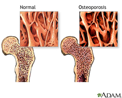 Osteoporosis is typically defined as a condition in which there is a persistent, net loss of both bone tissue and bone minerals due to an imbalance between the processes of bone formation and bone resorption. For instance, a relatively excessive blood concentration of the hormones which promote bone resorption (such as parathyroid hormone, cortisol, T3, and T4) can tip the scale toward the development of osteoporosis. Conversely, a relatively deficient blood concentration of the hormones which promote bone formation (such as HGH, IGF-1, estrogen, testosterone, calcitonin, and the active form of vitamin D) can tip the scale in the same direction. However, the etiology of osteoporosis is not unifactorial and is more complex than typically appreciated by physicians who simply prescribe hormone-replacement therapy or bisphosphonates for this condition (which is not a good idea, unless the patient is desiring to develop breast cancer, osteonecrosis of the jaw, or atrial fibrillation).
The interplay between parathyroid hormone, calcitonin, and the active form of vitamin D (1,25-dihydroxyvitamin D3) in the regulation of blood calcium levels is quite simple and well understood, so let’s focus on what is usually ignored or not well known regarding osteoporosis’ etiology. Firstly, gastrointestinal absorption of calcium is largely dependent upon a sufficient concentration of the active form of vitamin D, but “inorganic” versions of calcium (such as those found in most supplements) are very poorly absorbed and utilized regardless of vitamin D levels. Organic calcium salts (from plant-based supplements or whole foods) have been chelated or bound to a carbon-containing compound (like an amino acid or organic acid), which allows them to be more easily recognized and assimilated by the body. Conversely, the calcium present in pasteurized, homogenized milk is hardly assimilated by the human body as such milk is notably alkaline and lacks the enzymes needed for proper calcium absorption (calcium is absorbed best in an acid medium). So organically-grown vegetables (such as bok choy, cabbage, white carrots, turnip greens, mustard greens, almonds, broccoli, kale, and lima beans) are much better sources of usable calcium. Other issues which can impair the absorption of calcium include: pharmaceuticals used to treat acid reflux; surgical resection of some part of the GI tract; persistent use of glucocorticoid drugs (they promote urinary calcium excretion); and GI inflammation due to microbial infection, high sugar consumption, or gluten-related damage [1]. The second major factor that can be at play in the development of osteoporosis is the immune system’s hyperactivity due to intestinal hyperpermeability (or Leaky-gut syndrome). When business that shouldn’t be leaking out of the gut begins to, the immune system gets placed on high alert, and the adrenal glands and the liver can become notably weakened from having to respond to the onslaught of toxins/antigens. Such overactivity of the immune system can lead to an autoimmune response against osteoprotegerin (a glycoprotein which inhibits bone resorption), which can contribute to osteoporosis by increasing the rate at which bone is resorbed [2]. The toxins leaking out of the gut can also damage the hormone receptors embedded within the membranes of bone cells, so that the hormones which promote bone formation become less effective at doing so. The last factor we’ll discuss is strongly tied to the first two. To avoid going unnecessarily deep, the liver uses alkalizing minerals (such as calcium and sodium) to assist in its detoxification duties. When the liver is forced to deal with an onslaught of acidic toxins (from a leaky gut), it can begin protecting its sodium reserves, forcing the body to begin pulling calcium from the bones to buffer the acidic toxins [3]. Furthermore, an overly-taxed liver will be less efficient at recycling estrogen, which can forestall estrogen’s beneficial influence on bone formation (especially in women). A stressed liver can also pass on un-detoxed toxins to the kidneys, which can interfere with calcium reabsorption by the kidneys. Lastly, a leaky gut (and any excessive or persistent stress in general) can overstimulate the adrenal glands and their release of cortisol, which decreases intestinal absorption of calcium – this indirectly leads to a secretion of parathyroid hormone (which increases osteoclast or bone-resorbing activity). So you can see that simply prescribing estrogen, a calcium/vitamin D supplement, or bisphosphonates does not effectively correct the underlying progenitors in the development of osteoporosis. Of course, depending upon an individual’s specific etiology, a different resolution plan may need to be instituted, but I hope this article was helpful in illuminating the true origins of osteoporosis as conventional medicine’s typical understanding of this condition is embarrassing. References:
|
AuthorDenton Coleman is an Exercise Physiologist and Medical Researcher. Archives
October 2023
Categories |

 RSS Feed
